Suture-Less Mole Removal: Benefits, Process, and Recovery
Suture-Less Mole Removal: Benefits, Process, and Recovery
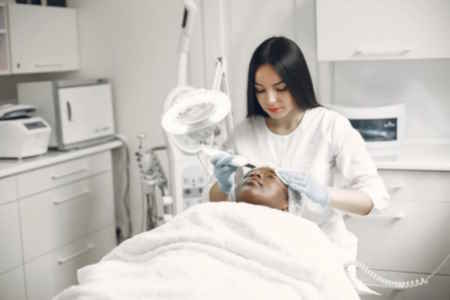
Mole removal can be performed for clinical assessment or appearance-focused reasons. Traditional excision may involve stitches and can leave a noticeable scar. By contrast, suture-less mole removal uses methods that typically do not require sutures. This approach may suit selected moles following clinical evaluation. A consultation with a qualified health professional is required to determine suitability and whether pathology testing is indicated.
Advantages of Suture-Less Mole Removal
1. Scarring Considerations
Suture-less techniques aim to minimise tissue disruption. Some people experience a finer or flatter mark compared with sutured excision, particularly for appropriately selected small or superficial lesions. However, any mole removal can leave a visible mark and outcomes vary based on skin type, mole size and depth, healing, and aftercare.
2. Recovery
These methods are generally less invasive than full-thickness excision. Many people resume usual activities soon after the appointment, depending on the method used and individual healing. Your clinician will provide specific instructions and discuss factors that could extend recovery.
3. Appearance-Focused Benefits
Suture-less removal is designed to blend the treated area with surrounding skin when appropriate for the lesion. Results differ from person to person, and a natural-looking finish cannot be guaranteed.
4. Convenience
Compared with surgical excision, suture-less methods are often completed in a single outpatient visit. Your clinician will advise if more than one visit or further care is needed.
Ideal Candidates for Suture-Less Mole Removal
1. Types of Moles
This approach is generally considered for benign lesions after assessment. If a mole is changing or has features that raise concern for skin cancer, a different technique and pathology testing may be recommended.
2. Health Considerations
Suture-less mole removal may be appropriate for people with good overall skin health who are seeking treatment for appearance-focused reasons. Certain medical conditions, medications, or active skin infections may affect suitability. Your practitioner will review your history and discuss options.
3. Other Factors
Suitability depends on clinical examination, including the mole’s size, depth, location, and the likelihood that pathology testing is required. Your clinician will explain the benefits and limitations of each method for your specific lesion.
Step-by-Step Process of Suture-Less Mole Removal
1. Initial Consultation
A clinician assesses the lesion to confirm that suture-less treatment is appropriate. Dermoscopic examination may be used. If there is any concern, excision with margins and pathology testing will be discussed.
2. Preparation
The area is cleaned. A local anaesthetic is usually used to improve comfort. The decision to use anaesthetic depends on the technique, lesion characteristics, and patient preference.
3. Removal Procedure
Suture-less mole removal may involve one or more of the following:
- Laser treatment: Energy targets the lesion tissue. Multiple sessions may be required depending on the lesion.
- Shave removal: The raised portion is carefully removed at or just below skin level.
- Radiofrequency ablation: Controlled energy removes tissue in thin layers to preserve surrounding skin.
The method is selected according to clinical findings and patient goals. Your clinician will outline likely outcomes, risks, and alternatives before proceeding.
4. Post-Procedure Care
After treatment, a protective dressing and topical care plan are provided. Written aftercare instructions explain how to keep the site clean and protected. Follow-up is arranged if clinically indicated.
Recovery and Aftercare
1. Healing Timeline
The area often forms a small crust in the first few days. This typically sheds within one to two weeks, although redness or colour change can persist for longer and may take weeks to months to settle. Some marks can remain.
2. Aftercare Tips
- Clean the area: Gently cleanse as directed to reduce contamination.
- Apply ointment: Use the recommended product to keep the site lightly moisturised.
- Protect from sun: Use clothing or sunscreen once intact skin has formed. Sun exposure during healing can increase pigment change.
- Avoid picking: Allow crusts to detach naturally to reduce irritation and scarring risk.
3. Follow-Up
Your clinician may recommend a review to assess healing or discuss any additional treatment. Seek advice promptly if you notice increasing pain, persistent redness, swelling, discharge, or any unexpected change.
Expected Results
When care instructions are followed, many people find the treated area blends with surrounding skin over time. Individual results vary and some textural or colour changes may remain visible. Your practitioner will discuss realistic outcomes based on your skin and the lesion treated.
Information on this page is general in nature, not a substitute for a consultation, and does not guarantee outcomes. Any examples or images reflect one case and may not predict results for others.
Risks and Recovery
All procedures carry risks. Your practitioner will explain these in a consultation and provide written information. Potential risks include bleeding, infection, delayed healing, discomfort, swelling, redness, crusting, changes in skin colour, sensitivity to sun, recurrence of the lesion, need for further treatment, scarring, and unsatisfactory cosmetic outcome. Rare complications can occur. Recovery experiences differ by individual and by method. Your clinician will provide tailored advice about activity restrictions, wound care, and when to return for review. If malignancy is suspected at any point, an alternative approach with pathology testing will be recommended.
“